Laboratory Equipment Details
Complete information about this device
Device Gallery
Available
Reservations
0Views
284Overall service quality rating
0/5
Information about devices and laboratories is clear and updated
0/5
The device search interface is effective
0/5
Steps to request the use of a device are clear
0/5
Devices are in good working condition
0/5
Contributes to obtaining accurate research results
0/5
Devices are available at the required times
0/5
Response to inquiries in a timely manner
0/5
Technical support is qualified
0/5
Contributed to the completion of research
0/5
Recommend your colleagues to use the service
0/5
Device Information
Device Name
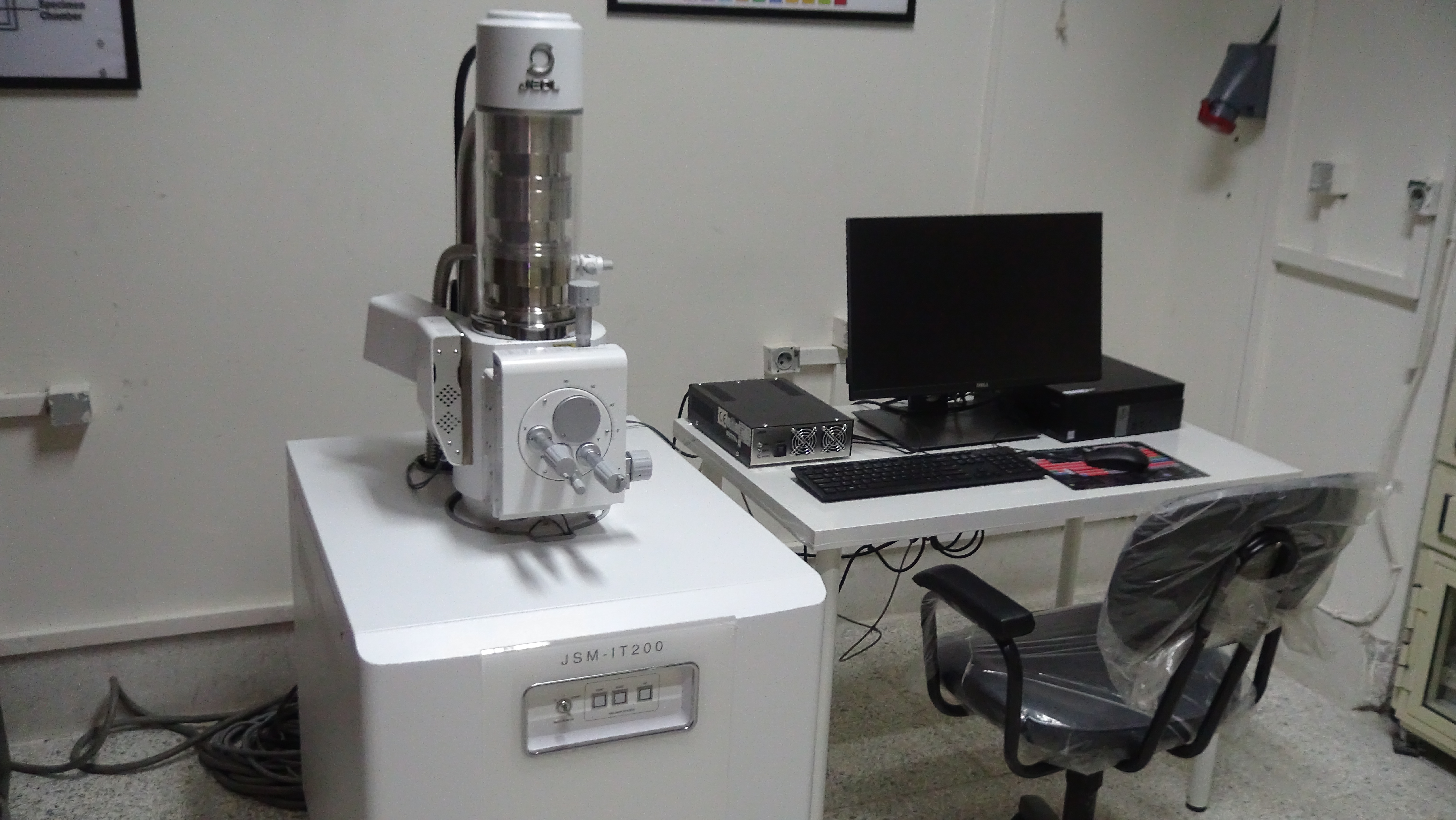
Scanning Electron Microscope (SEM)
Model
JEOL JSM IT200
Units Available
1
Manufacture Year
Manufacturer Website
Description
The scanning electron microscope has a magnification power of up to 300,000 times. It is used for imagining the surfaces of biological and non-biological samples. It provides high-quality and clear 3D images of nano samples. The device is equipped with an Energy Dispersive Spectroscopy (EDS) unit for analyzing different elements and determining the proportion of each element in the sample along with an illustrative diagram. Creating a map of the existing elements and their distribution.
Services & Pricing
| Service | Cost |
|---|---|
| Preparation and examination of a biological sample on the scanning electron microscope (SEM) + 5 images | 550/600 EGP |
| Preparation and examination of a non-biological sample on the scanning electron microscope (SEM) + 5 images | 500/550 EGP |
| Examination and analysis of a sample using EDX | 350/400 EGP |
| Sample analysis and creation of an elemental mapping | 1000/1100 EGP |
| Preparation and examination of a non-biological sample on the scanning electron microscope (SEM) + EDX + 5 images | 700/750 EGP |
| Preparation and examination of a non-biological sample on the scanning electron microscope (SEM) + Mapping + 5 images | 1250/1350 EGP |
| Sample analysis on the scanning electron microscope (SEM + EDX + Mapping) | 1350/1450 EGP |
| Taking extra images for any sample on the microscope (per image) | 40/50 EGP |